Languages
Label-Free Imaging
Advancing Cancer Research with Multiphoton Microscopy
Cancer is a major cause of death worldwide and, given the aging global population, the number of deaths due to cancer is expected to continue to increase. The relationships between cancer, immune cells, and the collagen matrix are complex, and analyzing them requires the implementation of new technologies and ideas. Understanding the mechanisms behind these relationships will enable the development of more precise therapeutics and faster diagnostic tools.
The application of MEM with label-free fluorescence lifetime imaging microscopy and second-harmonic generation microscopy helps to move cancer research forward by providing highly precise data about these relationships.
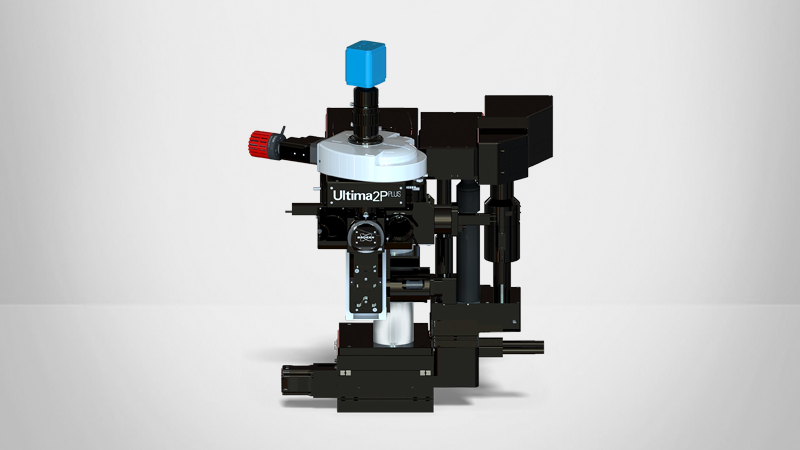
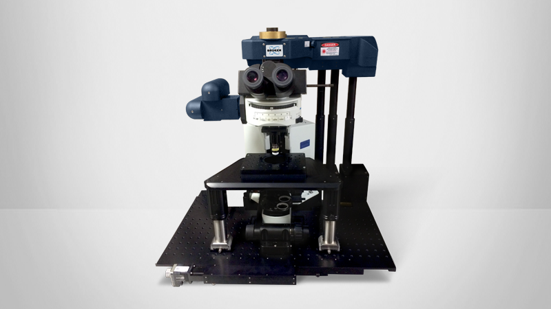
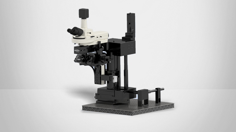
Explore Bruker MEM Technology
Over the past two decades, multiphoton excitation microscopy (MEM) has enabled detailed intravital imaging of metastasis in animal models of cancerwith supreme optical sectioning and minimal phototoxicity. In addition, MEM offers the possibility of fast imaging of unstained tissue based on its morpho-chemistry. As a result, MEM imaging has already greatly improved our ability to diagnose and treat cancer and will continue to play a significant role in preventing cancer-related deaths going forward.
Cancer researchers have successfully used Bruker multiphoton imaging systems to study the structure and physiological state of cells and tissues in normal and pathological conditions.
References
- 1. Datta R, Heaster TM, Sharick JT, Gillette AA, Skala MC. Fluorescence lifetime imaging microscopy: fundamentals and advances in instrumentation, analysis, and applications. J Biomed Opt. 2020 May;25(7):1-43. doi: 10.1117/1.JBO.25.7.071203. PMID: 32406215; PMCID: PMC7219965.
- 2. Walsh AJ, Mueller KP, Tweed K, Jones I, Walsh CM, Piscopo NJ, Niemi NM, Pagliarini DJ, Saha K, Skala MC. Classification of T-cell activation via autofluorescence lifetime imaging. Nat Biomed Eng. 2021 Jan;5(1):77-88. doi: 10.1038/s41551-020-0592-z. Epub 2020 Jul 27. PMID: 32719514; PMCID: PMC7854821.
- 3. Sagar MAK, Ouellette JN, Cheng KP, Williams JC, Watters JJ, Eliceiri KW. Microglia activation visualization via fluorescence lifetime imaging microscopy of intrinsically fluorescent metabolic cofactors. Neurophotonics. 2020 Jul;7(3):035003. doi: 10.1117/1.NPh.7.3.035003. Epub 2020 Aug 8. PMID: 32821772; PMCID: PMC7414793.
- 4. Szulczewski JM, Inman DR, Entenberg D, Ponik SM, Aguirre-Ghiso J, Castracane J, Condeelis J, Eliceiri KW, Keely PJ. In Vivo Visualization of Stromal Macrophages via label-free FLIM-based metabolite imaging. Sci Rep. 2016 May 25;6:25086. doi: 10.1038/srep25086. PMID: 27220760; PMCID: PMC4879594.
- 5. Keikhosravi A, Li B, Liu Y, Eliceiri KW. Intensity-based registration of bright-field and second-harmonic generation images of histopathology tissue sections. Biomed Opt Express. 2019 Dec 9;11(1):160-173. doi: 10.1364/BOE.11.000160. PMID: 32010507; PMCID: PMC6968755.


