Multi-mode PET technology provide new insights for clinical oncology research before
Positron emission tomography (PET) is a kind of widely used in nuclear medicine imaging technique that can be used to before the clinical diagnosis and clinical application.PET technology using radioactive tracer (radiotracers) provide the three-dimensional "functional imaging, display biomolecular activity in animal models and the spatial distribution in the human body.Before starting the clinical trials, small animal research and exploration in the preclinical phase it is necessary to verify imaging agent.
In this case, the PET imaging is increasingly in the stage of drug development, the researchers used because it provides data can infer to human studies in animal studies.Based on mechanisms of disease to provide important insights from molecules to the organ level, preclinical imaging is helpful to the development and evaluation of new treatment strategies.Small animal imaging deepened our understanding of disease development and potential therapeutic effect, and PET technology is driving the resources application in the clinical environment.
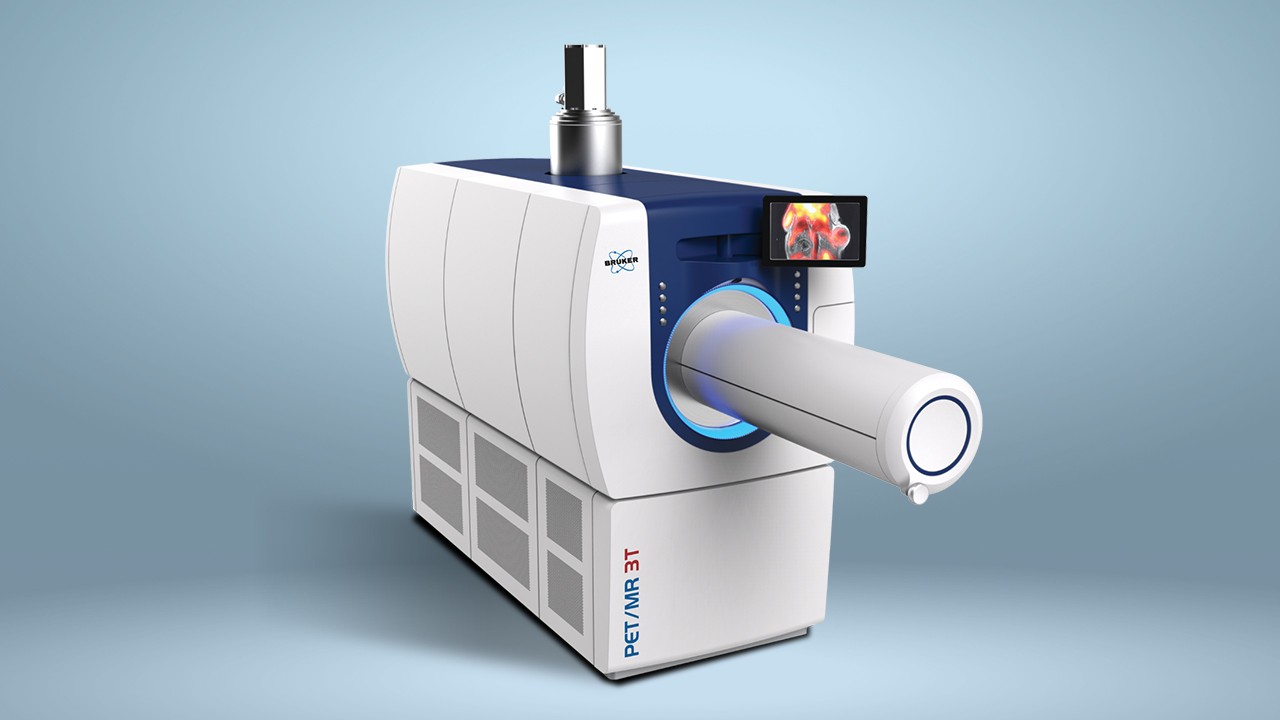
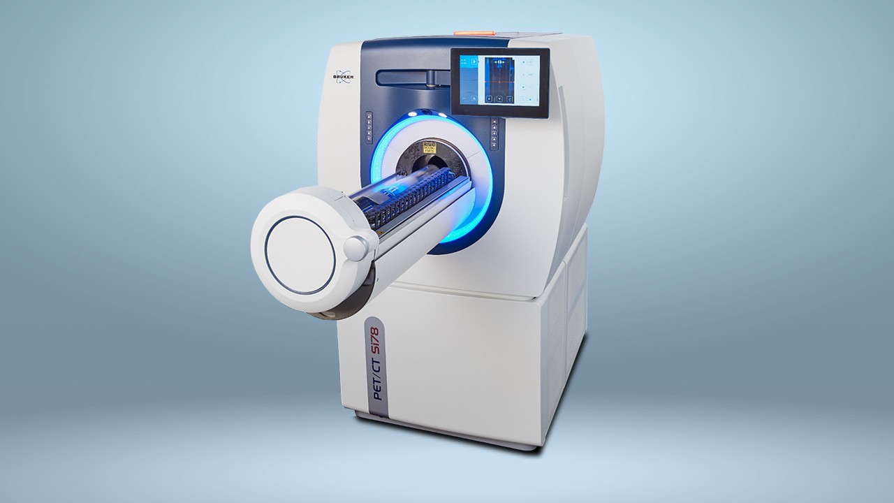
Provide the patients with personalized cancer therapy this demand, is pushing for preclinical research progress of PET in oncology.Many different types of cancer, including those who have not yet been detailed characterization of the tumor, and their different reaction to treatment, and to find new effective cancer therapy was a incredibly challenging.Non-invasive in vivo imaging techniques, such as PET, allowed the researchers to through real-time visualization cancer related process, a better understanding of the development of the tumor.The combination of PET and other imaging methods, such as magnetic resonance imaging (MRI), computed tomography (CT) and single photon emission computed tomography (SPECT), combination of structural and functional imaging in an experiment.PET/CT、PET/MR和PET/SPECT/CT多模态系统能够提供放射性核素、骨骼和软组织的定量三维断层图像,进一步增进了人们对癌症生物学和治疗的认识。
Download the complete application documents:


